Services
VITREO RETINAL SERVICES
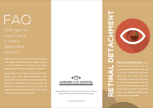
Retinal Detachment is the separation of the Retina from the underlying layers the line the inner walls of the eye. Through the Retinal Tear, liquid from the vitreous may pass through tear and detach the retina.
Vitreoretinal Services - Retinal Detachment
Retinal detachment is separation of the retina from the underlying layers that line the inner wall of the eye. Through the retinal tear, liquid from the vitreous may pass through the tear, and detach the retina. As the fluid accumulates, more and more of the retina detach. Detached retina loses its function; hence the person with retinal detachment loses vision suddenly or gradually.
Causes
The vitreous-a gel like material is present is maximum areas of eye. Meanwhile, the presences of retinal tear allow gel from vitreous space to pass through the hole and flow between the retina and the back wall of the eye. As a result the retina detaches from its underlying layer of support tissue at the back of the eye.
Symptoms
Most people notice floaters and flashes before the retina detaches. As the detachment increases a gradually enlarging dark shadow engulfs vision. It may appear as a curtain or a shade drawn slowly across the field of vision.
Retinal Detachment Treatment
Retinal tears with minimal or no detachment can be treated on an out-patient basis using laser therapy or cryopexy (freezing) procedures. These treatments decrease the risk of a retinal detachment. Retinal detachment may rarely occur even after these treatments; it is hence essential that the patient is on regular follow-up after the treatment.
Vitreoretinal Services - Diabetic Retinopathy
Diabetes is a disease that inhibits with the body’s ability to use and store sugar, which may lead to several health problems. Excess sugar present in the blood may cause damage in the entire body including eyes. Meanwhile, the circulatory system of the retina also gets attacked by diabetes. Diabetic Retinopathy is an eye complication that occurs in person due to Diabetes and which causes continuous harm to the retina. Moreover, it is the outcome of damage to the tiny blood vessels that feed the retina. These tiny blood vessels drip blood as well as other fluids that results in swelling of retinal tissue and clouding of vision.
Causes
Diabetic Retinopathy occurs mainly due to the changes in the blood vessels of the retina. Diabetes produces weakening of the blood vessels in the body.
Symptoms
Diabetic Retinopathy produces ocular symptoms only in the last stage of the disease. Only the best ophthalmologist in India can detect in early signs of Diabetic Retinopathy. So, all Diabetic patients should undergo yearly eye (Retinal) examination.
Treatment
Laser Treatment – Lasers are widely used in treating diabetic retinopathy and is performed as an out-patient procedure. In this treatment an intense and highly energetic beam of light is focused on the area to be treated with the aid of the slit lamp and a special contact lens. This will reduce the retinal thickening and stops bleeding. Additional treatment may be required depending on the patient’s condition.
Surgical Treatment – In some patients the vitreous may pull on the retina reducing vision severely. In such cases a surgical procedure called vitrectomy is performed. This surgery is done for advanced stage of Diabetic Retinopathy.
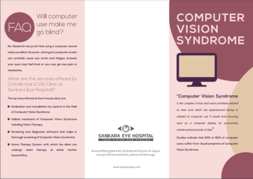
VISION THERAPY CLINIC – COMPUTER VISION SYNDROME
Our Vision Therapy centre includes facilities such as Computer Vision Clinic. Many aspects of computers and the work environment in which they are used are likely to cause or contribute to the development of eye or vision difficulties. Sankara Eye Hospital offers holistic, specialized treatment for Computer Vision Syndrome at the Computer Vision Clinic.
Causes
Regular use of computers for more than 3 hours in a day is known to predispose to Computer Vision Syndrome in the presence of one or a combination of the following factors:
- Uncorrected vision problems
- Poor lighting
- Improper viewing distances
- Glare from the computer screen
- Poor seating posture
- Back pain
Treatment
Treatment of Computer Vision Syndrome is achieved by
- Glasses: Normal or specially designed for computer eye.
- Vision Therapy: included various exercise of the eyes that help the eyes to focus, move and to work together.
- Eye Care: to prevent the recurrence of the symptoms of computer Vision Syndrome
- Artificial tears: to reduce dryness of eyes
- Altering ergonomics
Vision Therapy Clinic - Vision Rehabilitation
Vision is used in the completion of almost all daily activities. Loss of vision will require a period of training, adaptation and the learning of new skill. During this period the person is likely to require help from another person several times a day with a range of activities.
Vision Rehabilitation Provides
- Coping with vision loss
- Age appropriate milestones
- Independent daily living activities
- Guidance in selecting better model and method of education
- Travelling safety
- Taking care of the home
- Meeting career objectives
- Enjoying leisure activities
CATARACT
Cataract (motiyabind) is the opacity (clouding) of eye’s natural lens which is normally transparent. When lens of our eye becomes cloudy, light rays cannot pass through them easily and vision becomes blurred. It can develop in one eye or both eyes and has a variety of causes. The Cataract Clinics at Sankara are equipped with world class infrastructure and is overseen by the best ophthalmologists In India and paramedical staff. The clinics are equipped to screen, detect, diagnose and offer right procedural corrections for cataract. Sankara offers the world class eye care treatment at all 13 super speciality eye hospitals set up across 10 states of country .
What causes Cataract
Cataract development is a normal process of aging. Any person may develop it sooner or later. Cataract can also be present from birth. Researchers across the world have recognized factors that are associated with the formation as well as the development of cataract
Cataract Treatment
To a certain level, cataract affected vision can be corrected with prescription eye-glasses including contacts or bifocals. However, beyond a certain point prescription glasses may be ineffective and surgical options have to be considered. In a cataract surgery, the patients’ cloudy lens is removed and replaced with a transparent Intra Ocular Lens (IOL) to restore good quality vision.
Symptoms
At the front of the eye, there is a small space called the anterior chamber. Clear fluid flows in and out of the chamber to bathe and nourish nearby tissues. In Glaucoma there is an impairment in the dynamics of this fluid called aqueous humour due to varied causes, some of which are unknown leading to increase in intraocular pressure. Unless this pressure is controlled, it may cause damage to the optic nerve and loss of vision.
Treatment
At the front of the eye, there is a small space called the anterior chamber. Clear fluid flows in and out of the chamber to bathe and nourish nearby tissues. In Glaucoma there is an impairment in the dynamics of this fluid called aqueous humour due to varied causes, some of which are unknown leading to increase in intraocular pressure. Unless this pressure is controlled, it may cause damage to the optic nerve and loss of vision.
GLAUCOMA
Glaucoma is a group of eye diseases in which the retinal nerve fibres are damaged leading to progressive optic nerve damage resulting in decreased vision or even blindness. It is an important reason for poor quality of life due to loss of vision resulting in difficulty in near vision, working in dim light and driving.
What causes Glaucoma
At the front of the eye, there is a small space called the anterior chamber. Clear fluid flows in and out of the chamber to bathe and nourish nearby tissues. In Glaucoma there is an impairment in the dynamics of this fluid called aqueous humour due to varied causes, some of which are unknown leading to increase in intraocular pressure. Unless this pressure is controlled, it may cause damage to the optic nerve and loss of vision.
Diagnostic test for Glaucoma
Glaucoma can be easily diagnosed with simple and effective tests which can help in quantifying the anatomical as well as functional loss due to the disease. These tests are advised regularly during the follow up visits or when the disease is not under control and help in proper management of the patient.
Symptoms
Most types of Glaucoma cause no symptoms. Central vision stays normal until very late in the disease. If Glaucoma remains untreated, people may notice that although they see things clearly in front of them, they miss objects to the side and out of the corner of their eye. It seems as though they are looking through a tunnel.
Treatment
Glaucoma is one of the leading causes for loss of vision worldwide. However, this vision loss can be avoided by timely intervention, which is usually very simple in the form of medical treatment. Patients benefit from our state-of-the-art Glaucoma testing capabilities with specially trained Best Glaucoma specialists In India who are experts in diagnosing and providing either medical or surgical management for all types of Glaucoma. Although Glaucoma cannot be cured, it can usually be controlled.
LASIK AND PRK
At Sankara Eye Hospital, we understand the significance of clear vision and the impact it can have on your daily life. That’s why we are proud to offer state-of-the-art LASIK surgery, a revolutionary procedure that has helped millions of people achieve freedom from glasses and contact lenses.
LASIK and PRK are two distinct refractive surgery procedures aimed at correcting common vision problems, but they differ primarily in their approach to reshaping the cornea.
Lasik vs PRK
Recovery Time:
LASIK: The recovery time for LASIK is generally quicker. Patients often experience improved vision within a day or two as the corneal flap adheres rapidly, and discomfort is minimal.
PRK: PRK typically involves a longer recovery period as the epithelium needs time to regenerate.
Postoperative Discomfort:
LASIK: Patients undergoing LASIK often report minimal discomfort during the recovery period, with many experiencing clear vision relatively soon after the procedure.
PRK: PRK patients may experience some discomfort initially as the corneal surface heals. Pain and sensitivity to light are common in the first few days after PRK.
Corneal Thickness and Eligibility:
LASIK: It is generally more suitable for individuals with thicker corneas due to the creation of a corneal flap.
PRK: PRK may be recommended for individuals with thinner corneas as it does not involve creating a flap.
Suitability Factors:
LASIK: Often preferred for individuals seeking a quicker recovery and less postoperative discomfort.
PRK: Considered for those with specific corneal conditions or thinner corneas and for individuals where LASIK might not be suitable.
Ultimately, the choice between LASIK and PRK depends on individual factors such as magnitude of refractive error, type of refractive error, corneal thickness and lifestyle.
Why Choose Sankara for LASIK Surgery/PRK ?
Expertise and Experience
Sankara Eye Hospital is renowned for its expertise in eye care, backed by a team of highly skilled ophthalmologists and surgeons. Our LASIK/PRK specialists have years of experience in performing successful laser vision correction surgeries, making us a trusted choice for your vision enhancement needs.
Cutting-edge Technology
We invest in the latest and most advanced technology to ensure optimal outcomes for our patients. Our LASIK procedures are performed using cutting-edge laser systems that provide precision and accuracy, resulting in excellent visual outcomes.
Personalized Treatment Plans
No two eyes are the same, and at Sankara, we recognize the importance of personalized care. Our LASIK/PRK surgeons conduct thorough assessments to understand your unique eye anatomy, prescription, and lifestyle. This information is used to create a customized treatment plan tailored to your individual needs.
The LASIK Procedure at Sankara
Consultation and Evaluation
The LASIK journey begins with a comprehensive consultation and eye evaluation. Our experienced ophthalmologists will assess your eye health, discuss your visual goals, and determine your eligibility for LASIK surgery.
State-of-the-Art LASIK Technology
Sankara Eye Hospital utilizes US-FDA LASIK LASER Machines for LASIK procedures. This technology allows for precise corneal reshaping, resulting in improved vision and reduced dependence on glasses or contact lenses.
Comfortable and Quick Procedure
LASIK surgery at Sankara is a quick and virtually painless procedure. The majority of patients experience improved vision almost immediately, with minimal downtime.
Comprehensive Postoperative Care
Our commitment to your well-being extends beyond the surgery itself. Sankara provides thorough postoperative care to ensure a smooth recovery process and optimal visual outcomes. Our team is available to address any concerns and answer questions at every step of your LASIK journey.
Benefits of LASIK/PRK at Sankara Eye Hospital
Improved Vision: Experience clearer vision and reduced dependence on glasses or contact lenses.
Experienced Surgeons: Trust your eyes to a team of skilled and experienced LASIK/PRK surgeons.
Cutting-edge Technology: Benefit from the latest advancements in LASIK technology for precise and effective outcomes.
Personalized Care: Receive a customized treatment plan tailored to your unique eye characteristics and visual goals.
Comprehensive Support: From the initial consultation to postoperative care, we are committed to providing comprehensive support and guidance.
Schedule Your LASIK/PRK Consultation Today
Take the first step towards a life without visual limitations. Schedule a consultation with our LASIK/PRK specialists at Sankara Eye Hospital. Discover the freedom of clear vision and experience the world with newfound clarity.
Contact us today to embark on your LASIK/PRK journey at Sankara Eye Hospital – where your vision is our priority.
OCULAR ONCOLOGY
Retinoblastoma is a cancerous growth that occurs in the eye. The tumor is of two types. Retinoblastoma is a cancerous tumor needing immediate attention. Retinoblastoma can present as a white reflex at the pupil of the eye (Cat’s eye like appearance) or as a squint. Rarely if ignored in the early stage, it can cause protrusion of the eyeball.
What causes a Retinoblastoma
As in most cancers, we still do not know why some children develop Retinoblastoma. Retinoblastoma can however occur in families due to a genetic defect and children born in such families are definitely at risk
If either parent is affected, there is approximately 40% chance of future offspring developing Retinoblastoma.
Retinoblastoma Treatment
Removing the affected part so that it does not spread to other part of the body and cause death Using radiation treatment.
Giving intravenous medicines (chemotherapy).
Using laser or cryo treatment.
ORBIT & OCULOPLASTY
Oculoplasty is a discipline in ophthalmology that deals with plastic and reconstructive surgery of the peri-orbital and facial tissues which include the eyelids, orbit and lacrimal system.
Causes
Symptoms
Ptosis is the medical term for droopy eyelids. Droopy upper lids may be present in children since birth or present in the early years of life.
Orbit And Oculoplasty Treatment

PAEDIATRIC OPHTHALMOLOGY & STRABISMUS
Amblyopia is reduced vision in an eye that has not received adequate use during early childhood. Amblyopia, or “lazy eye,” has many causes. On the other hand, A Squint (Strabismus) is a condition of the eye that causes one of the eyes to turn inwards (converge), outwards (diverge) or sometimes upwards, while the other eye looks forward.
Amblyopia
Amblyopia is reduced vision in an eye that has not received adequate use during early childhood. Amblyopia, or “lazy eye,” has many causes. Most often it results from either a misalignment of a child’s eyes, such as crossed eyes, or a difference in image quality between the two eyes (one eye focusing better than the other). Amblyopia is the most common cause of visual impairment in childhood. The condition affects approximately 2 to 3 out of every 100 children.
Causes
Amblyopia mainly occurs during childhood when the nerve pathway from one eye to the brain does not develop adequately. Generally, when the eyes are not working together and send a wrong or blurred image to the brain, the brain gets confused and may start to ignore the image from the weaker eye resulting in amblyopia
Treatment
The goal of amblyopia treatment is to improve the visual acuity of the poorer eye, such that it’s acuity comes at par or within one or two lines of the good eye. With early diagnosis and treatment, the sight in the “lazy eye” can be restored. A recent NEI report suggests that older children can also benefit from amblyopia treatment.
Paediatric Ophthalmology & Strabismus – Squint
A Squint (Strabismus) is a condition of the eye that causes one of the eyes to turn inwards (converge), outwards (diverge) or sometimes upwards, while the other eye looks forward. The cause, severity, and direction of a squint vary from person to person. It is usually spotted in childhood, sometimes within weeks of a baby being born, and affects 5-8% of children (1-2 in every 30).
Causes
There are six muscles attached to the outside of each eye that are responsible for eye movements. When there is an imbalance of these muscles, a Squint occurs. The cause of the squint may not be obvious but can include a family history, long or short sightedness, injury or viral illness.
Symptoms
The most common symptom of a Squint is one of the eyes not looking straight ahead. In new-born babies it is quite normal for their eyes to ‘cross’ occasionally, particularly if they are tired. However, if you notice that this happens to your child beyond three months of age, it is advisable to talk to your ophthalmologist.
Starbismus Treatment
A Squint is a condition that should be treated as soon as possible after it’s detected. Treatment is most effective in very young children. A Squint will not disappear as the child gets older, and in fact the sight in the affected eye will gradually get worse.
UVEA
Uvea is the middle layer of eyeball. Variety of disorders affect the uvea, most common of them is uveitis. We have expert and well trained team of the best eye specialists In India and equipment to diagnose and treat uveitis.
Symptoms
Patients commonly experiences intermittent attacks of redness, pain and increased sensitivity to sunlight, decrease in vision.
Treatment
As uveitis is associated with other systemic conditions; eye specialist consults with other specialty doctors like chest physicians, rheumatologists, pathologist, and radiologists to investigate and treat this condition in best possible manner. For finding a cause; blood investigations and sometimes x-ray, CT scans are necessary. Sometimes, systemic drugs like steroids are necessary to treat.
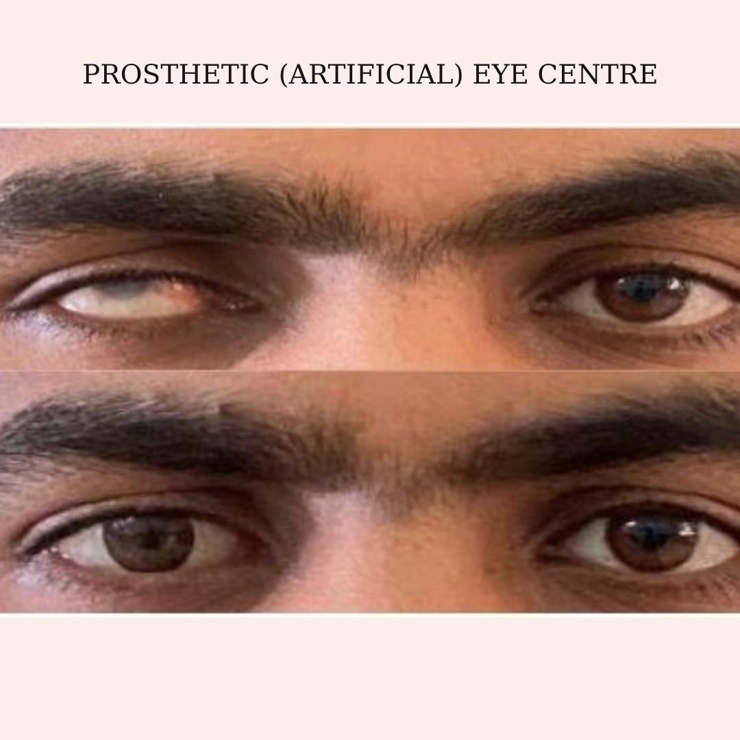
Prosthetic eyes are a very common cosmetic treatment for those who have lost an eye/or both to create a balanced facial appearance. This helps to improve your physical appearance. Lifts your self-esteem and confidence and visual aesthetic.Ocularistry @ Sankara is unique in our ability to provide both stock & customised prosthetic solutions to patients. The centre is a training & research hub looking to enhance the care to those requiring these services across India. Our patients have been from India, Sri Lanka, Bangladesh, Nepal, USA, Malaysia, Nigeria, Iran, Iraq, Yemen and many more.
Our Services
Customised Ocular Prosthesis: Discover a world of confidence and comfort with our meticulously crafted customised ocular prosthesis, providing a seamless and natural look.
Ready Made Prosthesis: Experience the convenience and immediate restoration of your appearance with readymade ocular prostheses.
Scleral Shells
Hollow Prosthesis
Modification/ Polishing of Prosthesis
How do we make it?
Through a combination of advanced techniques, we create customised ocular prostheses that look alike your natural eye. They are made using dimensionally stable, biocompatible material-PMMA.
We take 2-3 days to prepare customised prosthesis
Three-day procedure includes counselling, evaluation, measurement, hand painting the prosthesis, and fitting and polishing
The fitting of prosthesis is not at all painful.
Why Our clinic is unique?
Our team consists well experienced Oculoplasty consultants and expert team of ocularists.
Holistic approach to patient care with exceptional patient satisfaction
Comprehensive support including counselling to address the psychological aspects of wearing a prosthetic eye.
Eye Bank
Eye Bank
Corneal blindness is the fourth leading cause of blindness in the world. Corneal transplant is one of the primary treatment available for corneal blindness where damaged tissue is replaced with healthy donor eye tissue. Sankara Eye Hospital ventured into eye banking in the year 1985. Since then, it has opened 8 eye banks across various locations in India. Sankara has been actively engaged in eye donation awareness and cornea retrieval activities. The Eye Bank receives a pair of an eye almost every day and has collected over 19,262 eyes across all eye bank locations which are used for therapeutic and curative corneal transplant surgeries apart from research purpose. SEF, India also provides donor cornea to other eye banks for emergency transplants.
